The postmortem appearances, based on an incomplete postmortem examination, were those of liver failure after surgery. The final diagnosis was of Chloroform poisoning, this is most likely to have been subsequent to a prolonged anaesthesia in a debilitated sick patient.
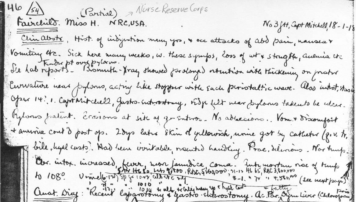
No 3 G H, Capt Mitchell 18-1-18
(Partial postmortem)
Fairchild Miss H. NRC,USA
Clinical Abstract), History of indigestion many yrs, and occasional
attacks of abdominal pain, nausea and vomiting etc. Sick here many weeks, with these symptoms,
loss of weight and strength, anemia etc.
Tender point over pylorus.
See lab reports - Bismuth Xray showed prolonged retention with thickening on greater curvature near pylorus, acting like stopper with each peristaltic wave. Also intestinal stasis.
Operation 14? Capt Mitchell, Gastro-enterostomy, ridge felt near pylorus taken to be ulcer. Pylorus patent. Erosions at site of gastro-enterostomy. No adhesions. Vomiting and discomfort and anuria continued post operatively. 2 days later skin of yellowish, urine got by catheter (g?lu ?glucose trace, Bile, hyaline casts.) Had been irritable, resented handling. Practically delirious. Normal temperature. Chronic intoxicated increased, fever, more jaundice, coma. Antemortem rise in temperature to 1080
Anatomical diagnosis Recent laparotomy and gastro-enterostomy. Acute parenchymal and fatty degeneration liver (Chloroform poisoning).
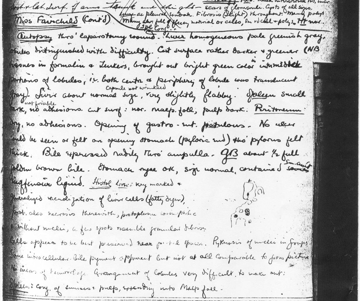
Autopsy thro' laparotomy wound.
Liver homogenous pale greenish gray. Lobules distinguished with difficulty. Cut surface raather darker and greener (NB tissues in formalin ?Zenkers (a tissue stain) brought out bright green color in intermediate portions of lobules, ie. Both center and periphery of lobule was translucent gray.) Liver about normal size, Capsule not wrinkled. Very slightly flabby.
Spleen small, dark, not friable, no adhesions. Cut surface: normal malpighian follicles, pulp dark.
Peritoneum dry, no adhesions. Opening of gastro-enterostomy patulous. No ulcer could be seen or felt on opening stomach (pyloric end) tho' pylorus felt thick. Bile expressed readily thro' ampulla.
GB (Gall Bladder) about half full golden brown bile. Stomach rugae OK. Size normal, contained small amount offensive liquid.
Histology Very marked and generalized vacuolation of liver cells (fatty degeneration). Probably also necrosis therewith. Protoplasm (?) without nuclei, a few spots resemble granular ?fibrosis (?debris). Cells appear to be best preserved near (?) spaces. Pyknosis of nuclei in groups. Some intracellular bile pigment apparent but not at all comparable to gross picture. No areas of hemorrhage. Arrangement of lobules very difficult to make out.
Spleen Congestion of sinuses and pulp expanding into Malpighian follicles.
![]()